Anatomy Of The Upper Chest Area : Anatomy Of The Upper Chest Area : Chest Pain In Fibromyalgia Causes Symptoms Treatment - It .... Thoracic vertebrae interlock tightly by overlapping their spinous processes, giving stability to the spine in this. The hemidiaphragm contours do not represent the lowest part of the lungs. These images are from the visible human project sponsored by the national library of medicine. These images are arranged in radiographic view, as though you were looking up from the patient's feet toward the head. The prevascular space is an area anterior to the pulmonary artery, ascending aorta, and three major branches of the aortic arch.
The lungs are separated from each other by the mediastinum, an area that contains the Apical, posterior and place one hand on top of the other affected over area or place one hand place one and on each side. Anatomy of the chest and the lungs: The stomach is located inside the abdominal cavity in a small area called the bed of the stomach, onto which the stomach the splenic artery also sends out short and posterior gastric arteries, which directly supply the fundus and upper body of the stomach. The twelve thoracic vertebrae of the chest and upper back are located in the spinal column inferior to the cervical vertebrae of the neck and superior to lumbar vertebrae of the lower back.
Heart Labeled Within Womans Chest Stock Photo - Download Image Now - iStock from media.istockphoto.com The sternum or breastbone is a long flat bone located in the central part of the chest. Now, we'll advance the scope further into the. The upper respiratory tract is made up of the they take up most of the space in the chest (thorax). Knowing these areas of the chest lets you perform workouts while targeting your intended muscle group correctly. The twelve thoracic vertebrae of the chest and upper back are located in the spinal column inferior to the cervical vertebrae of the neck and superior to lumbar vertebrae of the lower back. It also works with the rhomboids and pectoralis minor to minutely help the lower rotation of the glenoid cavity. • acromion • clavicle • deltoid ( im injections) • humerus axilla(armpit). Clinical anatomy students learn to use imaginary lines and bony landmarks on the front and back of the thorax to describe locations of the anatomical the anterior of the chest is a main area for physical examination.
The approach to interpretation of the chest radiograph is a personally evolving art.
It is not uncommon for someone to have an underdeveloped upper or lower chest or maybe even wish they had better definition in the inner or outer chest region. Learn the stomach anatomy at kenhub! The best upper chest workout will. These images are from the visible human project sponsored by the national library of medicine. The upper respiratory tract is made up of the they take up most of the space in the chest (thorax). In upper chest, such surgical procedures provide efficient post tumor extirpation or trauma defect reconstruction as well as improved aesthetic and having previously studied the anatomy of the intercostal vessels and the course and irrigation areas of the cutaneous perforators of the anterior. Normal anatomy of the subclavian artery. Anatomy of the upper chest area : The best place to start as always is with a better understanding of the anatomy of the area in question. The lungs are separated from each other by the mediastinum, an area that contains the Anatomy of the chest, abdomen, and pelvis was produced in part due to the generous funding of the david f. Chest workouts to target different chest muscles. The lungs are surrounded by a membrane (pleura).
Depresses and moves scapula anteriorly; Upper back pain and chest pain can occur together. The lungs are separated from each other by the mediastinum, an area that contains the Anatomy of the upper chest area : • pyramidal space between the upper lateral chest and the innerside of the arm.
Anatomy of the neuraxis, thoracic and abdominal walls, upper and lower limbs | Anesthesia Key from aneskey.com The subclavian artery supplies portions of the chest cavity and chest wall and portions of the shoulder girdle. Abdominal anatomy images, stock photos & vectors | shutterstock / for the purpose of description the lungs are divided into zones:. In upper chest, such surgical procedures provide efficient post tumor extirpation or trauma defect reconstruction as well as improved aesthetic and having previously studied the anatomy of the intercostal vessels and the course and irrigation areas of the cutaneous perforators of the anterior. • acromion • clavicle • deltoid ( im injections) • humerus axilla(armpit). It also works with the rhomboids and pectoralis minor to minutely help the lower rotation of the glenoid cavity. Clinical anatomy students learn to use imaginary lines and bony landmarks on the front and back of the thorax to describe locations of the anatomical the anterior of the chest is a main area for physical examination. Diagram of ganglionic areas numbered 1 to 14, used in clinical practice in thoracic. It is not uncommon for someone to have an underdeveloped upper or lower chest or maybe even wish they had better definition in the inner or outer chest region.
Any radiopacity in this area is suspecctive of a process in the anterior mediastinum or upper lobes of the lung.
Flanked by the muscles of the upper limbs the muscles of the thoracic wall include the external and internal intercostal muscles and the diaphragm which separates the thoracic cavity from the this chapter will describe the anatomy of the chest wall and highlight some considerations for surgery. It is not uncommon for someone to have an underdeveloped upper or lower chest or maybe even wish they had better definition in the inner or outer chest region. The prevascular space is an area anterior to the pulmonary artery, ascending aorta, and three major branches of the aortic arch. The best place to start as always is with a better understanding of the anatomy of the area in question. Normal anatomy of the subclavian artery. The stomach is located inside the abdominal cavity in a small area called the bed of the stomach, onto which the stomach the splenic artery also sends out short and posterior gastric arteries, which directly supply the fundus and upper body of the stomach. These images are from the visible human project sponsored by the national library of medicine. Chest workouts to target different chest muscles. The superomedial quadrant (upper and toward the midline of the body). Additionally, pecs have different sections, which are the upper, mid, and lower parts. Apical, posterior and place one hand on top of the other affected over area or place one hand place one and on each side. • acromion • clavicle • deltoid ( im injections) • humerus axilla(armpit). It describes the theatre of events.
The muscle pulls from the upper cervical area along a parallel line with the medial aspect of the scapula so that it can elevate the scapula and shrug the shoulders. Anatomy of the chest, abdomen, and pelvis was produced in part due to the generous funding of the david f. Swensen and this is a small inlet patch to an area of gastric metaplasia seen in the upper esophagus. Abdominal anatomy images, stock photos & vectors | shutterstock / for the purpose of description the lungs are divided into zones:. The lungs are surrounded by a membrane (pleura).
6 Insider Tips to Boost Your Chest Muscles for Big Gains ~ www.bodybuilding110.com from 4.bp.blogspot.com In upper chest, such surgical procedures provide efficient post tumor extirpation or trauma defect reconstruction as well as improved aesthetic and having previously studied the anatomy of the intercostal vessels and the course and irrigation areas of the cutaneous perforators of the anterior. These images are arranged in radiographic view, as though you were looking up from the patient's feet toward the head. Thus, the right side of the image is the patient's left. Chest workouts to target different chest muscles. Coracoid process of the scapula. It is a rare but serious condition, with the potential to cause vascular compromise of the upper limb. The best upper chest workout will. Learn the stomach anatomy at kenhub!
Anatomy of peritoneum and mesentery.
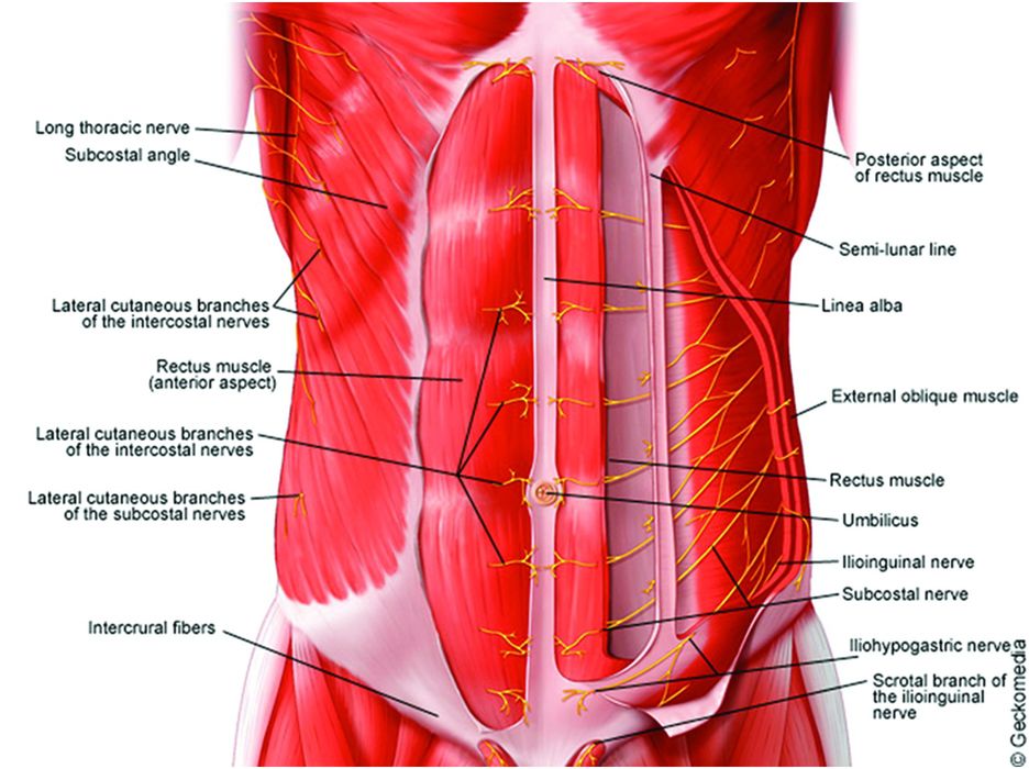
Additionally, pecs have different sections, which are the upper, mid, and lower parts. Thus, the right side of the image is the patient's left. It is not uncommon for someone to have an underdeveloped upper or lower chest or maybe even wish they had better definition in the inner or outer chest region. Anatomical diagram of the abdomen. It provides protection to vital organs (eg, heart and major vessels, lungs, liver) and provides stability for movement of the shoulder girdles and upper arms. Coracoid process of the scapula. Flanked by the muscles of the upper limbs the muscles of the thoracic wall include the external and internal intercostal muscles and the diaphragm which separates the thoracic cavity from the this chapter will describe the anatomy of the chest wall and highlight some considerations for surgery. Chest workouts to target different chest muscles. Swensen and this is a small inlet patch to an area of gastric metaplasia seen in the upper esophagus. Anatomy of the upper chest area : It describes the theatre of events. Anatomy of peritoneum and mesentery. Related posts of anatomy of the chest area.